
Bleeding when you brush, bad breath that keeps coming back, or gums that look “puffy” are not just small annoyances. They are often the first signs of periodontitis — an infection of the tissues that hold your teeth in place. The good news: with early diagnosis and careful, microscope-assisted treatment, we can stop the disease from progressing, protect bone, and keep your own teeth stable for the long term.
Microscope dentistry is the heart of our approach. Magnification and focused light let us see plaque and calculus that the naked eye misses, especially subgingival (below gumline) deposits. That extra visibility means more complete cleaning with less trauma to healthy tissue and a smoother recovery.
What periodontitis is (and why it matters)
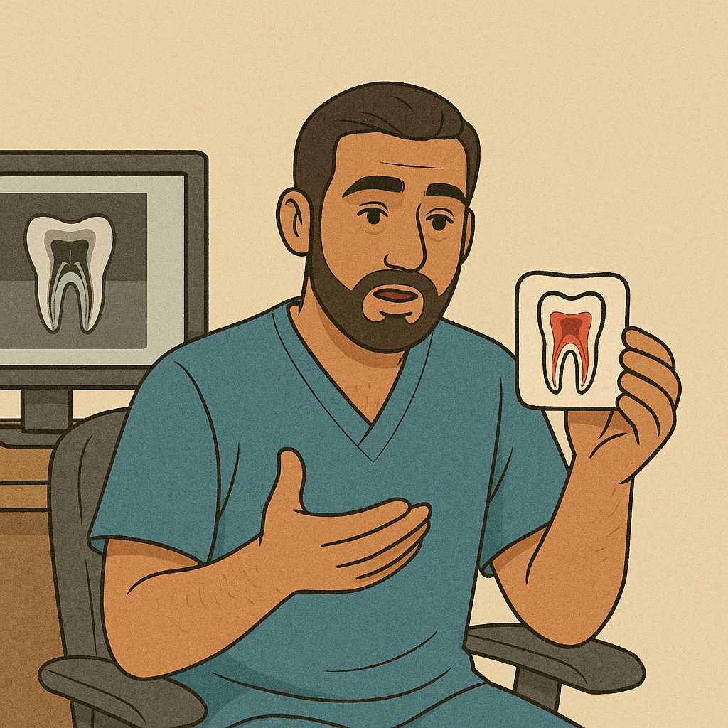
Periodontitis is long-standing inflammation of the periodontium — the gum, ligament, and bone that support your teeth. It usually starts with plaque that hardens into calculus. The attachment between tooth and gum loosens, pockets form, and bacteria move deeper. Without treatment, bone slowly recedes and teeth loosen.
You might hear different names:
- Gingivitis: early, reversible gum inflammation.
- Mild periodontitis: early bone loss with shallow pockets.
- Moderate or advanced periodontitis: deeper pockets, gum recession, tooth mobility.
- Apical / periapical periodontitis: infection at the tip of a tooth’s root (often linked to a nerve problem) that may exist together with gum disease.
If you notice bleeding, a metallic taste, or gaps that trap food, it is time for a periodontal exam. Periodontitis is a leading cause of tooth loss in adults, but it is treatable and controllable at any stage.
How we diagnose — step by step
Your first visit is a structured, calm evaluation:
- Periodontal charting pocket by pocket, with bleeding and mobility scores.
- Digital X-rays (and CBCT only when it genuinely changes decisions) to map bone and root anatomy.
- Microscope-assisted exploration to identify hidden plaque and calculus before we start.
- Risk profile (smoking, diabetes, clenching, medications, family history).
- Plan and sequencing — from non-surgical care to re-evaluation, and advanced treatment only if pockets remain deep.
You will see your measurements and photos. We explain them in simple language and agree on a plan that fits your schedule.
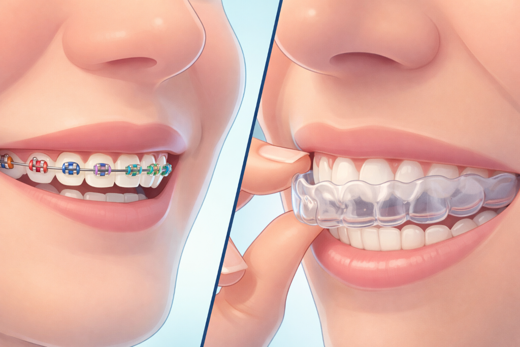
Non-surgical treatments (most people start here)
Think of this as a deep, precise clean — not a quick polish.
- Scaling and root planing (SRP): under local anaesthetic we remove plaque and calculus from the root surface and smooth micro-irregularities so bacteria cannot easily reattach. The microscope helps us be thorough and gentle.
- Local antimicrobials: in selected deep pockets we place targeted gels or rinses to reduce bacterial load.
- Adjunctive laser therapy: a gentle laser helps disinfect inflamed tissue. It is an addition to mechanical cleaning, not a replacement.
- Bite care and splinting: if teeth are mobile, we can adjust the bite and, when helpful, splint teeth together while gums heal.
- Systemic support: advice on smoking cessation, diabetes control, and dry-mouth management — all of which affect healing.
Re-evaluation is usually at 6–8 weeks. Many patients stabilize at this stage. If deep pockets persist, we discuss targeted advanced options.
Surgical and advanced options (for stubborn deep pockets)
When infection sites remain after excellent non-surgical care, we treat them precisely:
- Minimally invasive flap surgery: to access complex root surfaces, remove tenacious calculus, and contour bone for easier cleaning.
- Regenerative procedures: in specific vertical bone defects we use membranes and grafts to encourage the body to rebuild support (guided tissue regeneration, enamel matrix derivatives, or bone grafts).
- Mucogingival surgery: for gum recession with sensitivity or aesthetics concerns, a connective-tissue graft can improve coverage and thickness.
- Combined endodontic–periodontal care: if you have apical periodontitis (infection at the root tip), we coordinate root canal retreatment or apical microsurgery. The sequence is important: we treat the cause first, then the consequences.
Costs and insurance — clear, written, and tailored
We keep fees transparent and personalised, because no two mouths are the same.
Before and after — what to expect
Right after treatment: mild tenderness, temporary sensitivity to cold, and light bleeding the first day. A soft brush, careful flossing/interdental brushes, and the mouthrinse we prescribe help things settle quickly.
Weeks 1–8: bleeding reduces, breath improves, teeth feel smoother, and pockets start to shrink as swelling subsides. We re-measure at your review and decide whether you are ready for maintenance or need targeted advanced therapy.
Long term: with good home care and regular professional maintenance, results are stable for years. The aim is control, not “once and for all” — just like blood pressure or diabetes.
Choosing the right clinician for gum treatment
When you search periodontitis treatment near me, look for:
- Use of magnification or a microscope during cleaning and surgery.
- Full periodontal charting shared with you.
- A re-evaluation step before suggesting surgery.
- Clear coordination with endodontics for periapical or chronic apical periodontitis treatment if needed.
- A realistic maintenance schedule and education on home care.
At TRUE SMILE, we treat gums with the same respect as teeth — because healthy gums are what make every filling, crown, or implant last.
Prevention and long-term maintenance
- Professional hygiene: usually every 3–4 months for the first year, then tailored to your risk.
- Home routine: electric toothbrush technique, properly sized interdental brushes, and a nightguard if you clench.
- Lifestyle: reduce sugar frequency, quit smoking, manage reflux and dry mouth with your physician where relevant.
- Early signals: call us if you notice bleeding, a new food trap, or sensitivity that lasts more than a few days.
Consistent maintenance is the single best way to prevent periodontitis from getting worse.